Insights on site
Competence area musculoskeletal diagnostics
In one of its most important areas of competence – musculoskeletal diagnostics – DIE RADIOLOGIE combines knowledge, experience and a wide variety of examination options at several locations.
Cross-site area of competence
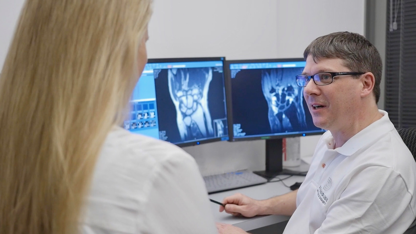
The diagnosis of diseases and injuries of the musculoskeletal system using MRI has been one of the main focuses of our practice for over 25 years. In order to respond individually to the complaints of our patients, we now operate different magnetic resonance tomographs at several locations, from dedicated joint scanners to semi-open MRI devices to high-field 3-Tesla MRI devices of the very latest generation.
Examination of the musculoskeletal system
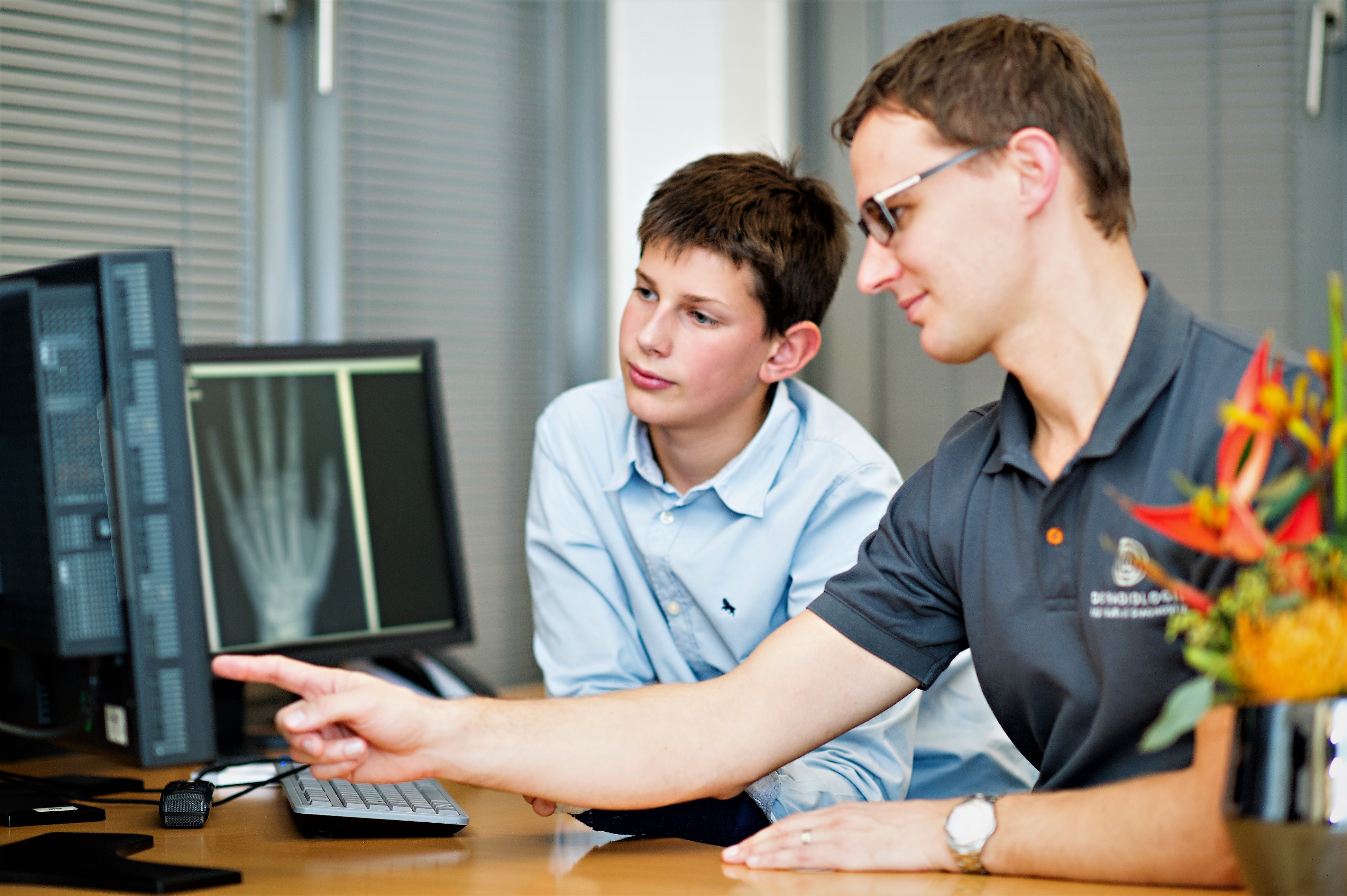
Musculoskeletal diagnostics includes diagnostics of the skeleton, joints, ligaments and muscles. In order to respond individually to the complaints of the musculoskeletal system, we now operate different magnetic resonance tomographs, from dedicated joint scanners to semi-open MRI devices to high-field 3-Tesla MRI devices of the very latest generation. As radiologists, we continue to see ourselves as X-ray doctors, in many radiological practices this is no longer a given. However, classic X-ray imaging is still the primary diagnostic for many questions. Today as a digital process with the lowest possible exposure to radiation. Our modern multislice computed tomography devices are the further development of X-ray diagnostics. The high-resolution cross-sectional imaging of the skeletal system allows three-dimensional images to be created and even the smallest changes in the skeleton to be recognized.
Special examinations
- direct MR or CT arthography: the examination of different joints with the administration of a contrast agent for certain questions
- MRI examinations of very small joints with the special joint scanner
- Bone density measurement using the DEXA method in cases of suspected osteoporosis
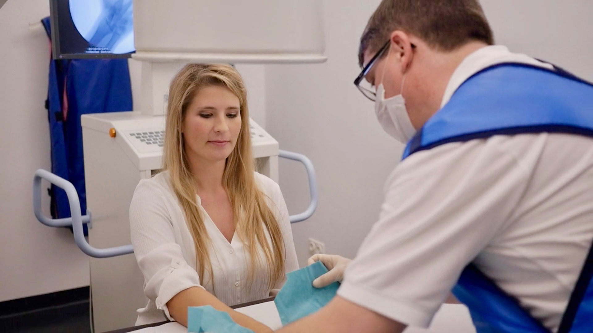
In direct arthrography, MRI or sometimes also CT examination of a joint is performed after a contrast agent has been injected into the joint. With the aid of this technology, which is indicated for special issues, primarily structures of the joint capsule and the cartilaginous parts of a joint can be assessed more accurately than in conventional MRI. This technology is used most frequently for the hip joint, the shoulder, and the wrist, sometimes also on the ankle.
The contrast agent is injected into the joint with a thin needle, about as big as one used for blood sampling, under sterile conditions and after local anaesthesia under X-ray control. Afterwards, the actual imaging is performed in the MRI machine.
The following risks are possible but very rare results of joint puncture: infection, bleeding or an allergic reaction to the contrast agent or the local anaesthetic.
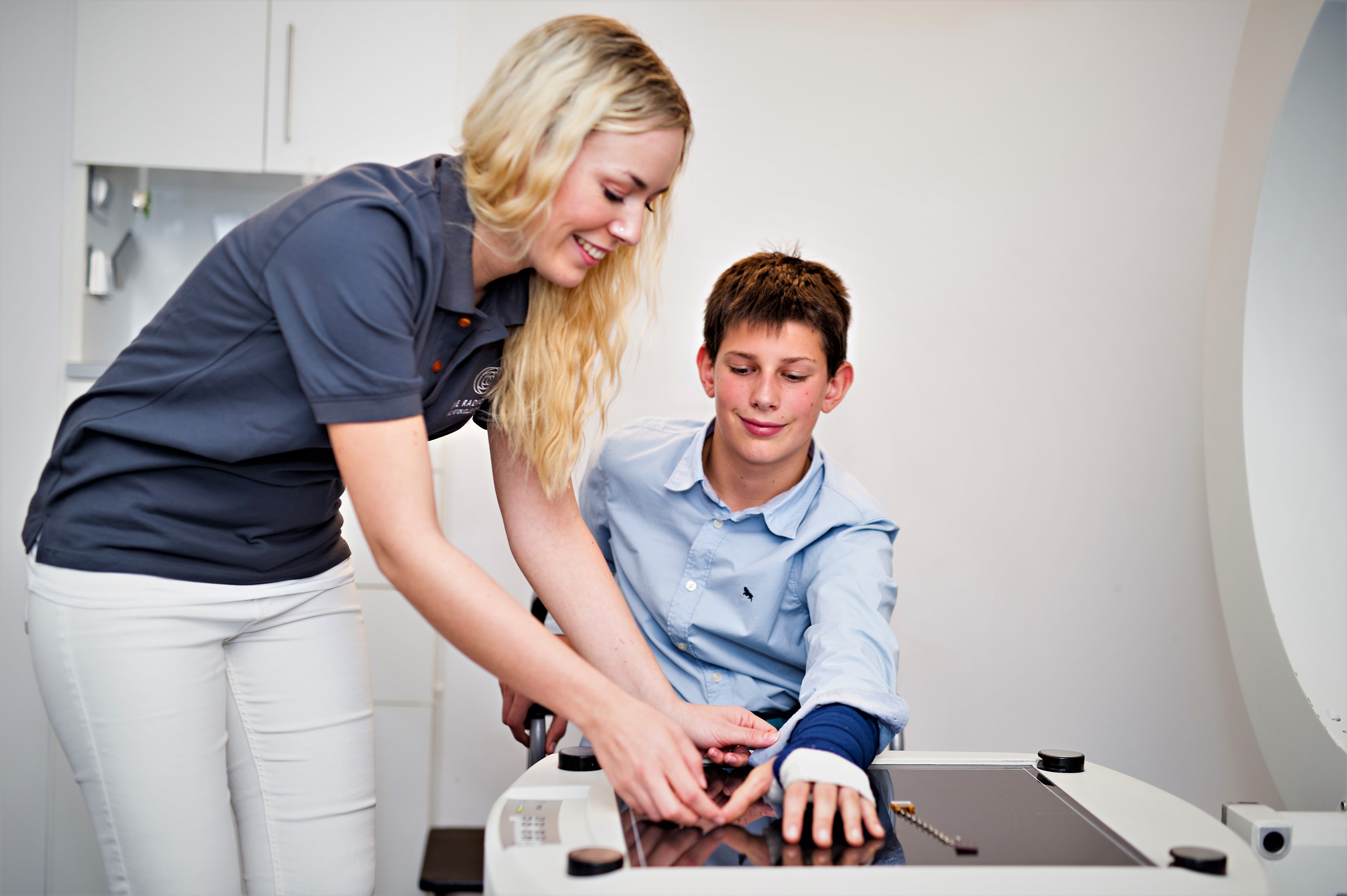
Our Heimeranplatz practice is equipped with a small, very powerful MRI scanner (joint scanner) for examinations of the hand/wrist and foot/ankle. During the examination, the patient sits comfortably next to the machine and only their hand or foot is inside the tunnel of the machine.
Osteoporosis is one of the most frequent diseases in ageing people. During bone metabolism, bone is increasingly lost, bone mass diminishes and the bone microstructure crucial for stability is changed. As a result of this, bone stability decreases, and the risk of bone fracture increases particularly in the spine and the thigh. However, increased bone loss can also occur as a result of medication intake (e.g. cortisone, some medications for breast cancer treatment).
The consequences of a fracture with osteoporosis are often serious and can have a detrimental effect on the quality of life, culminating in care dependency.
Today osteoporosis is a worldwide health problem. The WHO (World Health Organisation) has classified osteoporosis as one of the ten main widespread diseases.
The only way to diagnose osteoporosis at an early stage is to measure bone density. For this purpose, DEXA measurement (dual-energy X-ray absorptiometry) is usually performed, during which the bone density of the lumbar spine and the femoral neck is measured with very weak X-rays. Because the X-ray radiation is so weak, the 5 to 10 minute examination takes longer than a normal radiograph. DEXA measurement is the standard method for diagnosing and evaluating the progress of osteoporosis recognised and recommended by the WHO and the DVO (German Umbrella Association for Osteology). We offer DEXA measurement at our München Zentrum facility.
According to current DVO (Dachverband Osteologie – German Umbrella Association for Osteology) guidelines, bone density measurement is generally recommend as a basic diagnostic tool for women aged 60 and over and for men aged 70 and over. If risk factors such as diabetes mellitus type 1 or rheumatoid arthritis exist or if certain medications are taken, DEXA measurement is recommended for women already after menopause and for men aged 60 and over. For further information about bone density measurement, go to our examination bone density measurement.
Another method for measuring bone density is a QCT examination. This special (quantitative) computed tomography for measuring bone density is performed only in exceptional cases if DEXA measurement is no longer conclusive due to severe degenerative changes in the spine.
For osteoporosis prophylaxis, it is advisable to maintain not only a healthy lifestyle including exercise but also a healthy diet with sufficient intake of calcium and vitamin D3. The specific drug therapy partially depends on the cause of osteoporosis; please consult your attending physician about this.
Control examinations of bone density should not be performed at intervals shorter than one year.
Concentrated knowledge and experience
Comprehensive exchange of information in diagnostics Musculoskeletal imaging is a core area of radiological imaging and is performed by all of our doctors. In complex or unclear cases, however, there is always an exchange with our team of experts for musculoskeletal imaging: we are also always in close contact with our colleagues from orthopedics and trauma surgery.
We are here for you.
Continuous education for your health! In order to be able to offer you the best possible diagnostics, our experts regularly deepen their knowledge of musculoskeletal imaging by participating in national and international committees and congresses.
We are a certified training practice
As recognition of our competence and commitment to musculoskeletal diagnostics, our practice was certified by the German Society for Musculoskeletal Radiology (DGMSR) for the training of musculoskeletal experts in early 2016. The training is carried out by the instructors Prof. Dr. Mike Notohamiprodjo and Dr. Armin Seifarth.
Appointment and Contact
Our reception team will be glad to help you with all organisational questions. We are further happy to answer your medical questions – before and after the examination.
Get in touch with us: by phone, by making an appointment online or if you have any questions via our contact form.
*For our online appointments we are using a service of the company Doctolib GmbH, Berlin.