Locations
Munich:
Lucile-Grahn-Straße
From practice

Downloads
Competence area prostate diagnostics
With the multimodal imaging of the prostate MRI, DIE RADIOLOGIE offers the currently best method for imaging the prostate and for the exclusion or detection of a tumor.
Prevention is better than cure
Early detection is essential! Prostate imaging is becoming increasingly important in preventive care, treatment decisions and aftercare: with 50,000 new cases every year, prostate carcinoma is the most common malignant tumor in men. As with many other types of cancer, what causes it to develop is largely unknown. Genetic factors, a high-fat diet, environmental factors and hormonal influences are likely to be the cause. For example, the risk of developing the disease is about 4 to 5 times higher if the father or brothers have prostate cancer.
Multimodal imaging and multiparametric examination via MRI
More precise diagnoses through a combination of methods: MRI offers greater diagnostic certainty thanks to multimodal imaging. Perfusion, diffusion imaging and, if necessary, MR spectroscopy provide precise information about the location and spread of a possible tumor without having to intervene in the body.
Increased hit rate
Prostate MRI is superior to conventional PSA, palpation, and untargeted biopsy procedures alone. The results of an MRI examination can be extremely helpful to your treating urologist for a biopsy – the multiparametric MRI increases the hit rate.
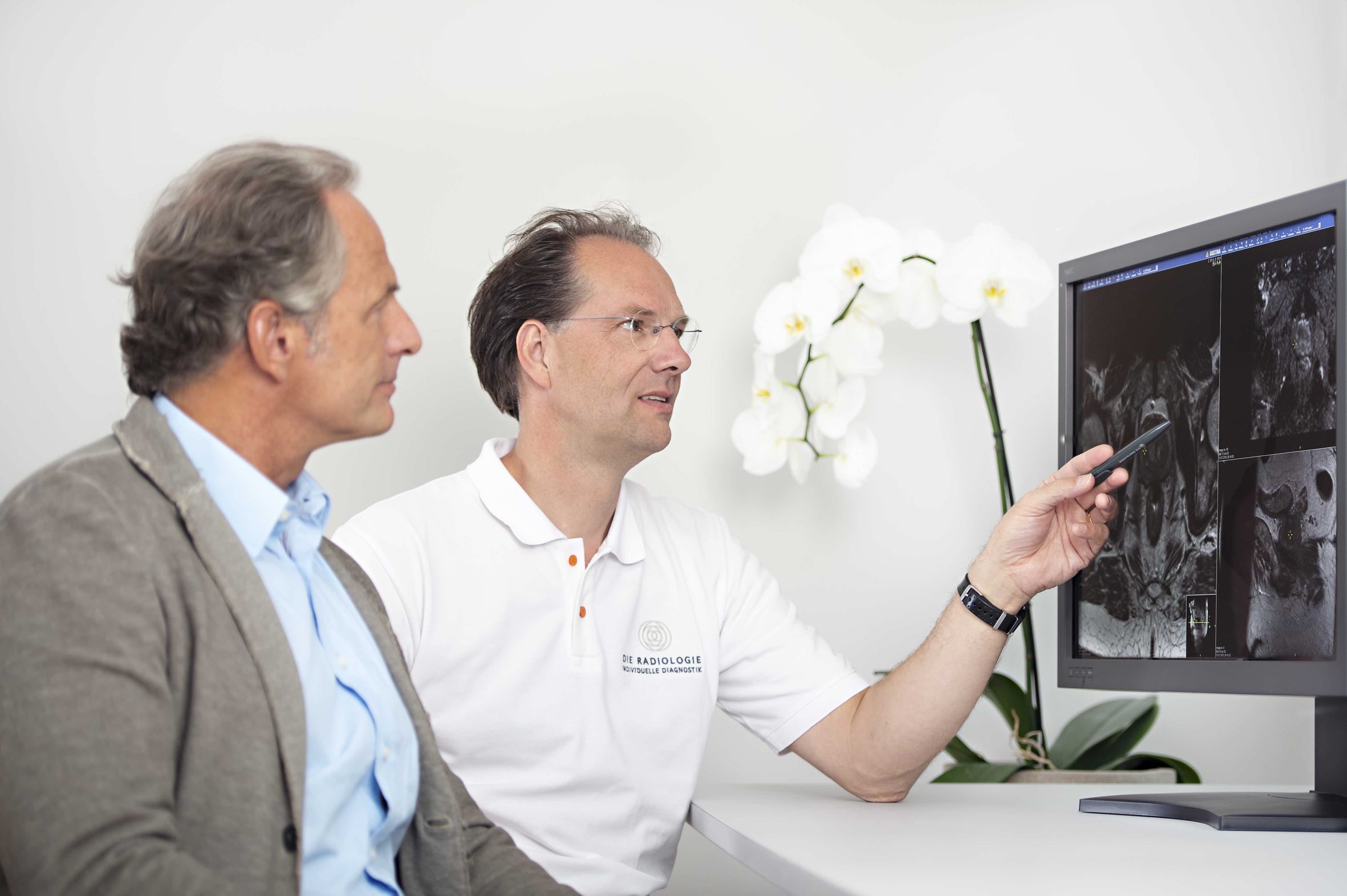
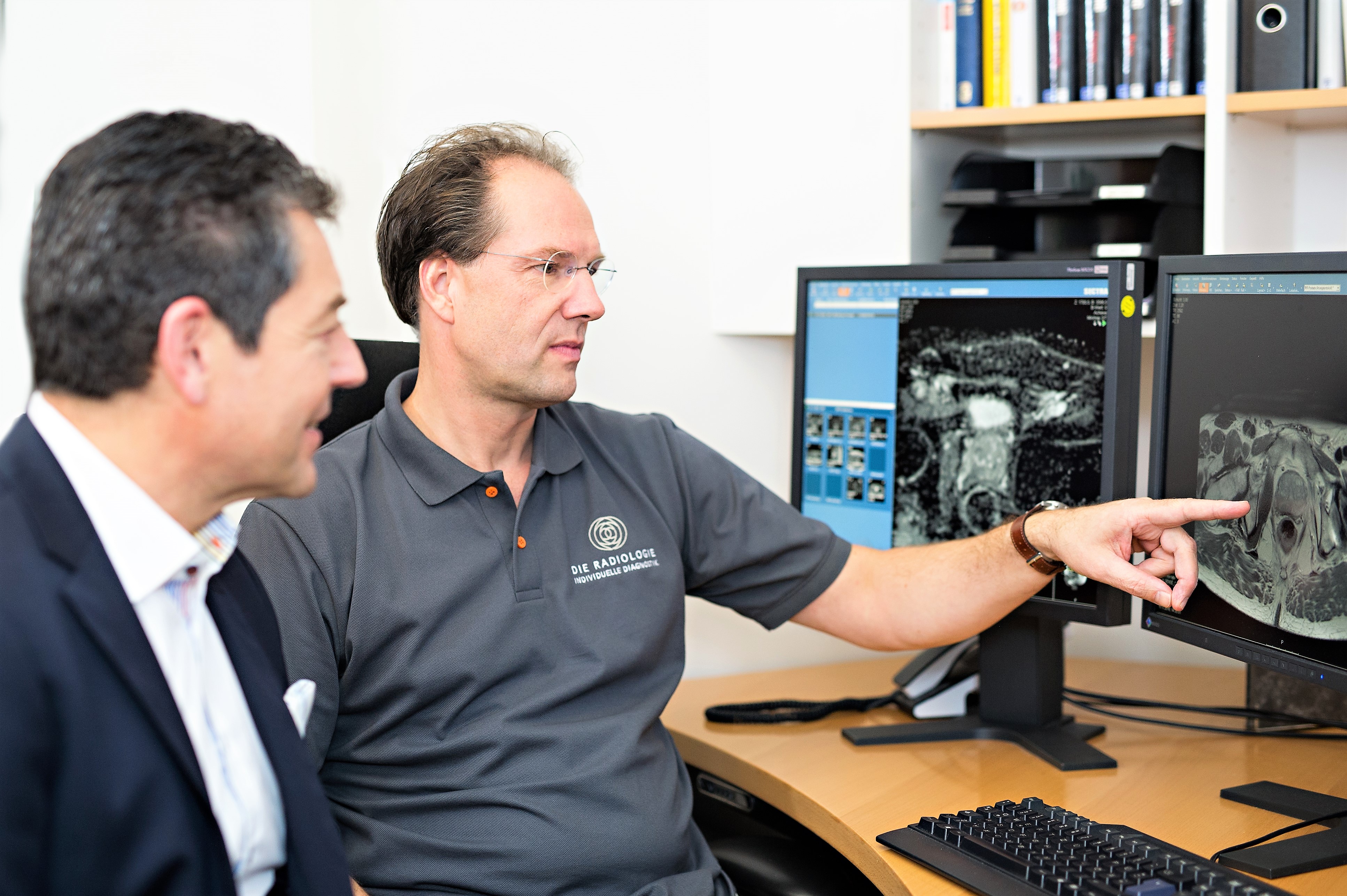
Concentrated knowledge and experience
Prostate diagnostics are primarily carried out on our modern 3-Tesla MRT devices in Lucile-Grahn-Strasse, in Rosenheim and Starnberg. Our experts, who are certified in the field of prostate MRI, take the time to discuss the examination procedure and the examination results with you.
Additional information
In the following you will find further important information on the assumption of costs, the indication for MRI as part of screening and in the event of suspected prostate cancer. Also more information on the multimodal approach of the study.
Palpation of the prostate recommended from the age of 50 is unfortunately not very reliable. Particularly small tumours are hard to detect with this method. Transrectal sonography is also conclusive only to a limited extent. Therefore, urologists determine the so-called PSA value (prostate-specific antigen) in addition. If it exceeds 4.0 ng/ml, there is suspicion of a tumour, and a tissue sample (biopsy) of the prostate should be taken. The reason: About 30% of 50-year-old men and up to 70% of over 80-year-olds have asymptomatic prostate cancer – often hidden amongst benign tumours.
However, the PSA value is high in over half of the cases with benign and harmless prostate diseases. Usually random biopsy (up to 18 samples) achieves a hit rate of only 30 to 40%. Thus this procedure is far from ideal.
If no tumour cells are found in a biopsy, that still does not rule out a tumour by any means. Then the biopsy has to be repeated if necessary. If a tumour is detected, it often remains unclear how extensive or aggressive it is.
For diagnosis of prostate cancer, magnetic resonance imaging is clearly superior to other imaging procedures. Thanks to the possibility of functional MRI diagnostics, it is currently the best imaging method for depicting the prostate and provides important information about the exact location and spread of a tumour as well as its aggressiveness in a non-invasive procedure. MRI can help to prevent possibly unnecessary diagnostic and therapeutic interventions for less aggressive tumours or benign changes.
Suspicious areas can be identified by MRI before a planned biopsy (tissue removal) significantly increasing the probability of finding the tumour. If a biopsy is necessary, the image data obtained during the MRI examination can be transferred directly to the urologist’s ultrasound images and thus mark the (sonographically usually invisible) tumour for the biopsy (so-called MRI fusion biopsy).
Suspicious areas in the prostate have to be biopsied to enable accurate classification and to confirm the diagnosis. MRI fusion biopsy is a novel procedure which enables the detection of particularly small and inconveniently situated tumour lesions with considerably higher accuracy (up to 85%) by combining the MRI scan with sonography during the biopsy.
If a tumour is detected (incl. in the biopsy), MRI can provide valuable information for planning treatment, for example, whether surgery or radiation is more appropriate or whether it is possible to wait and see (a technique known as ‘active surveillance’). For not every tumour grows aggressively.
Safety and certainty for patients can be significantly improved with prostate MRI since aggressive tumour growth can already be detected at an early stage.
Since magnetic resonance imaging does not require any X-rays, it is also perfectly suited to further monitoring in the case of unclear findings.
MRI is also instrumental in finding a so-called local recurrence (renewed PSA increase after therapy).
Conversely, aggressive tumour growth is relatively improbable if prostate MRI does not reveal any tumours.
1. High-resolution imaging
High-resolution MRI pictures of the prostate and its surroundings show the exact anatomy as well as suspicious areas (arrow: small prostate carcinoma).
2. Diffusion-weighted imaging (DWI)
Another innovation of prostate imaging that we offer at our surgery is so-called diffusion imaging, in which the diffusion (motion) of water molecules is examined and displayed graphically. Particularly aggressive prostate cancer can be detected more easily this way since water diffusion is limited by the increased cell density in the tumour.
3. Perfusion imaging (dynamic MRI)
During a perfusion examination, the contrast uptake in the prostate is examined over time as an indicator of blood circulation. Since tumours need nutrients for growth, they are usually supplied increasingly with blood, which distinguishes them from normal prostate tissue.
4. MR spectroscopy (MRS)
Additional accuracy thanks to high field MRI spectroscopy (MRS) of the prostate
With this special procedure, a tumour can be directly detected by suspicious metabolic products. In prostate cancer, citrate levels typical of normal prostate tissue are low and choline levels are high. During the examination, the prostate is systematically scanned for such suspicious areas.
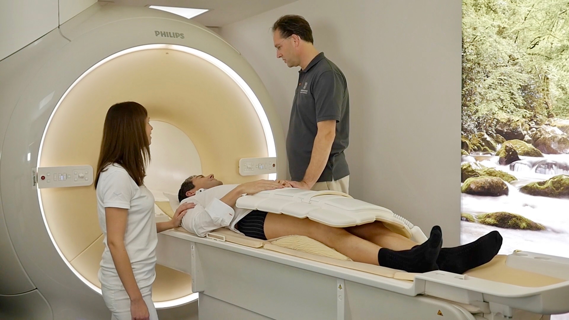
The magnetic resonance scanner creates high-resolution images of the prostate and also precisely depicts surrounding structures such as the seminal vesicles and the pelvic floor. In conventional MRI systems with a field strength of maximally 1.5T, it is usually necessary to insert an endorectal coil for this purpose. An unpleasant procedure which we spare our patients by using a state-of-the-art 3T high field MRI scanner with multi-transmit technology.
The entire examination is performed without a coil while you are lying down comfortably and takes approx. 25 minutes.
To visualise the blood circulation of the prostate as well as lymph nodes and to enable better differentiation of other structures, a well tolerated contrast agent (gadolinium) is administered intravenously in an arm. Please inform us, if you have a relevant allergy or any kidney dysfunction. During the examination, the urinary bladder should be as empty as possible.
Since prostate MRI reacts very sensitively to motions, well tolerated medication which suppresses bowel movement is administered before the examination (Buscopan). However, you should drink as little as possible and avoid tea and coffee before the examination. Gassy foods (e.g. beans) must be avoided on the day before the examination.
Important: an MRI examination of the prostate should not be performed during the first 6 weeks after a prostate biopsy.
Private health insurance providers in Germany usually reimburse the full screening examination costs. Patients insured under state/government insurance plans in Germany can have prostate MRI performed as a self-payer service. Unfortunately, cost coverage is currently not provided for this elaborate examination by compulsory health insurance funds.
We are here for you.
Our experts in the competence area for prostate diagnostics are at your disposal for questions with detailed and comprehensive advice, precise examinations and precise diagnoses. Benefit from our many years of experience in prostate diagnostics. On request, we can also offer you a second diagnosis or second opinion.
Appointment and Contact
Our reception team will be glad to help you with all organisational questions. We are further happy to answer your medical questions – before and after the examination.
Get in touch with us: by phone, by making an appointment online or if you have any questions via our contact form.
*For our online appointments we are using a service of the company Doctolib GmbH, Berlin.