Locations for this examination:
Munich:
Abdominal organs and thyroid:
München Zentrum
Mammary sonography:
Schwabing
Prinzregentenplatz
Surrounding Area:
Rosenheim
Mammary sonography:
Fürstenfeldbruck
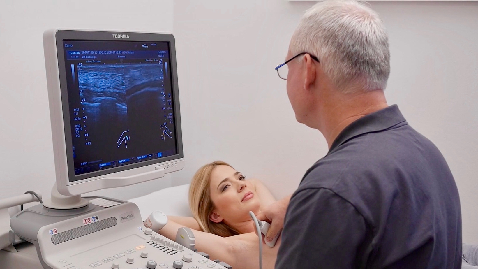
Ultrasound diagnostics
DIE RADIOLOGIE offers sonographic examinations for ultrasound diagnostics in its group practice. The technical principle behind a sonographic examination is relatively simple: an ultrasound probe emits sound waves which it receives as a reflection from the different deep layers of the body. The computer calculates an image of the inside of the body from the time difference between the transmitted and received sound and displays it on a screen.
We are here for you.
At our locations, our friendly and competent team of doctors and assistants ensure that this completely painless routine examination runs quickly and smoothly – we look forward to taking care of you!
Information on ultrasound examinations (sonography):
- no X-rays are used
- Visually guided puncture is possible for examinations of the breast and thyroid gland.
Most examinations take place while lying down, some examinations are also carried out while sitting.
The transducer is moved over the part of the body to be examined. The area is then examined from different directions and positions. The organs can be displayed simultaneously on one screen. The doctor can print out images using a video printer and store them digitally in our image archiving system.
There must be no air between the transducer and the skin, otherwise the sound waves cannot penetrate the body. In order to obtain an interference-free image, a neutral gel is applied to the skin and the transducer as a contact medium.
After the examination, we will discuss the result with you and, if necessary, advise you on further measures. You will receive the imagery right away. The actual report is sent directly to the referring colleague.
When examining the abdomen, the image quality is severely affected by air and the contents of the intestine. For this reason, you should not eat anything that causes flatulence (pulses, vegetables, salad, cabbage) the day before the examination. The examination should be carried out as sober as possible. On the day of the examination, you should therefore abstain from eating or drinking anything until the sonography.
When examining other body regions such as the neck, mammary glands or connective tissue, there are no special features to consider.
Which organs can be examined?
With the help of sonography, the structure of various organs and body regions can be examined easily and completely painlessly.
Sonography is mainly used in our practices:
- for assessing the organs in the upper and lower abdomen
- to examine the throat including the thyroid gland
- to examine the female breast
Sonography is also a valuable imaging method for showing enlarged lymph nodes, examining soft tissue (muscles and ligaments) and joint structures, and examining blood vessels.
Sonography is not suitable for examining air-filled organs such as the lungs or the gastrointestinal tract. On the other hand, organs filled with fluid, such as the gallbladder, can be visualized well by ultrasound.
We offer sonography for the following:
- the abdominal organs
- the chest
- the thyroid
- the musculoskeletal system and soft tissue
The upper abdominal sonography is used to clarify various diseases:
- Unclear upper abdominal complaints
- Liver: for example fatty liver or cysts
- Gallbladder: for example gallbladder/bile duct stones, gallbladder polyps
- Pancreas: for example pancreatitis
- Spleen: for example spleen enlargement
- Aorta (large abdominal artery): for example aortic aneurysm
- Kidney: for example urinary obstruction, kidney stones
- Lymph nodes: for example enlargements
Thyroid sonography is used for:
- Thyroid sizing
- Examination of nodules in the thyroid gland
- Evaluation of thyroid dysfunction
- Control after thyroid surgery or radioiodine therapy
- Monitoring after thyroid hormone therapy or iodide therapy
The breast is also referred to as "mamma" in medical jargon. This is why the ultrasound examination of the breast is also called “mammary sonography”.
Mammasonography is used for:
- Distinguishing between cysts and nodules
- Examination of nodes (benign or malignant criteria)
- Diagnosis of breast cancer in dense mammary gland tissue that is difficult to examine with mammography
- Confirmation of a suspicious finding found in mammography
- Diagnosis of nodules in young women
Appointment and Contact
Our reception team will be glad to help you with all organisational questions. We are further happy to answer your medical questions – before and after the examination.
Get in touch with us: by phone, by making an appointment online or if you have any questions via our contact form.
*For our online appointments we are using a service of the company Doctolib GmbH, Berlin.