Locations for this examination:
Munich:
München Zentrum
Am ISAR Klinikum
Nuclear medicine examinations
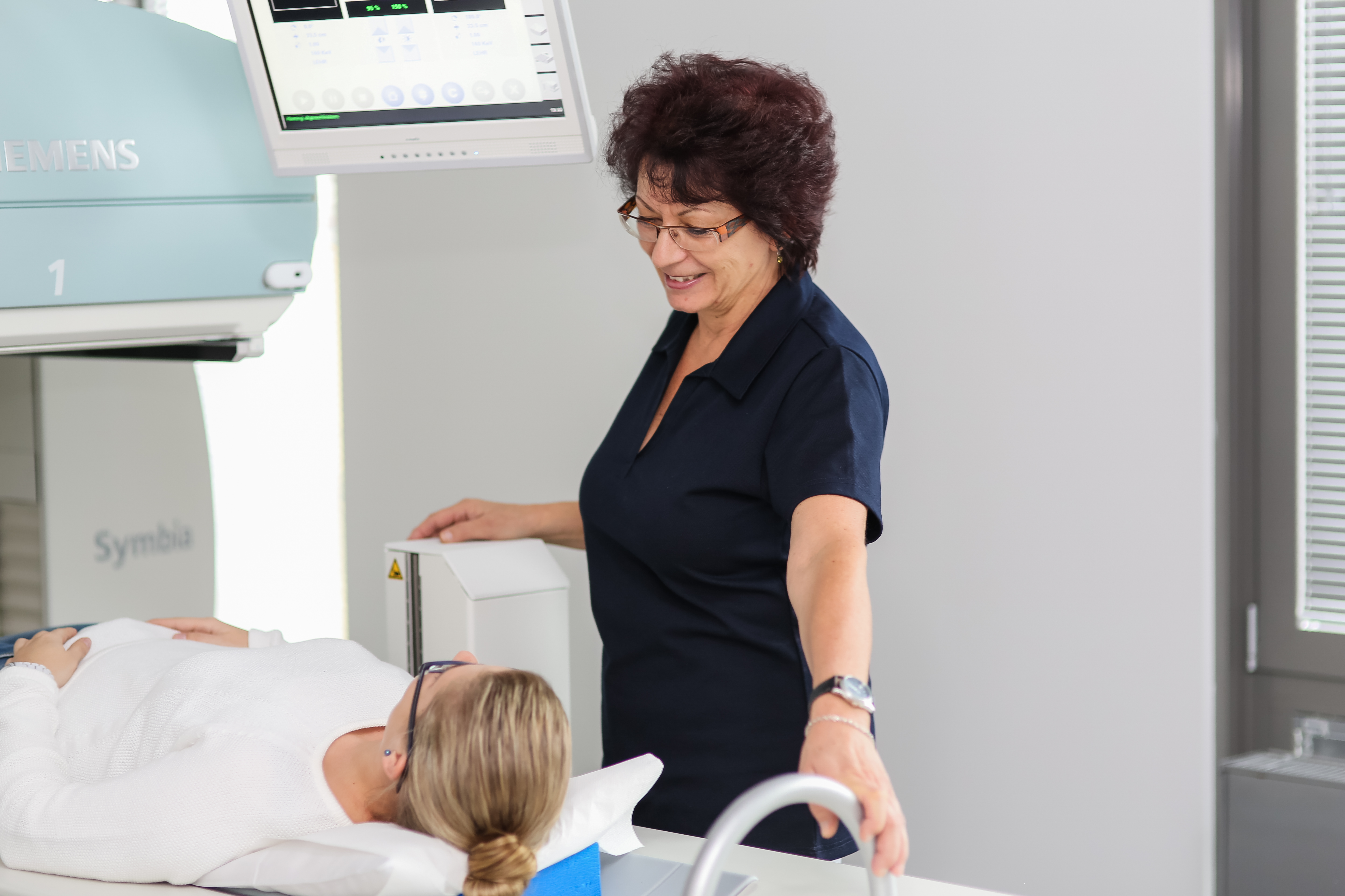
DIE RADIOLOGIE also offers nuclear medicine examinations at the various locations of the radiological practice network. These enable a functional diagnosis of the metabolism of the organs and the surrounding tissue – which is particularly important when assessing the condition of the thyroid gland. However, nuclear medicine methods are also used to diagnose the skeleton or the heart.
We are here for you.
At our locations for nuclear medical examinations, we take care of you efficiently and comprehensively. Our friendly and competent team of doctors and assistants ensure smooth processes – from reception to the examination to the explanation of the findings.
Our examinations
- Thyroid scintigraphy
- Bone scintigraphy
- Myocardial scintigraphy
- Renal function scintigraphy
- Thyroid puncture
Procedure and side effects
Depending on the examination, specific contrast agents are administered via the vein. After a certain waiting time, the accumulation and distribution of the contrast agent in the body is recorded with modern double-head cameras. These enable fast and precise imaging and examination. Side effects due to the extremely low radiation exposure are not to be expected.
Normally, no special preparation is required for a nuclear medicine examination. However, depending on the type of examination, you may have to remain sober (e.g. in the case of myocardial scintigraphy). In individual cases it may also be possible that you have to take certain medications before the examination or have to leave out medication your usually are required to take. Sometimes we also need additional information, e.g. the result of a blood test (e.g. thyroid scintigraphy) or previous X-ray examinations (e.g. question about loosening of a joint prosthesis in skeletal scintigraphy or question about pulmonary embolism in pulmonary perfusion scintigraphy). You will be informed about the details when you register for the examination or we will clarify this directly with your referring doctor.
During a scintigraphy, you will usually be given a small amount of a weakly radioactive substance in a vein in your arm. This weakly radioactive substance usually consists of the actual emitter (usually technetium 99m, abbreviated: Tc99m) and – depending on the organ to be examined and the medical case – a specific molecule to which the emitter is coupled.
The radioactive substance is distributed in the body via the bloodstream and accumulates in the organs to be examined. The radioactive signals can often be recorded by a gamma camera just a few minutes after administration (e.g. thyroid scintigraphy). Sometimes, however, a longer waiting period is necessary so that the substance can accumulate long enough (e.g. skeletal scintigraphy). Depending on the examination, the recording lasts between 5 and 45 minutes and takes place either lying down, sitting or standing. After the examination, the evaluation is carried out on the computer.
You are in no way affected by the nuclear medicine examination. The low-level radioactive substances used are usually without side effects, usually decompose very quickly or are quickly excreted through the kidneys. There are usually no allergies.
In some cases, additional examinations such as ultrasound or blood tests are necessary.
Examination duration: the examinations can vary greatly depending on the organ to be examined: from 30 minutes to four hours in several recordings at different times. After the examination, we will discuss the result with you and, if necessary, further measures. You will receive the imagery for the referring doctor at the same time. The findings are usually sent separately to the doctor treating you.
Nuclear medicine examinations are usually carried out to assess the function of organs and tissues. Such dysfunctions can often be the first sign of an illness.
Nuclear medicine examinations are not in competition with other imaging examinations, but a useful addition to the diagnostic process. They can be used to detect certain diseases earlier and more reliably. Many pathological changes can already be discovered with nuclear medicine examinations when other examination methods do not yet reveal any indications of a health problem. Examples are the disease of the coronary arteries and thus the impending heart attack or the cause of bone pain.
The most common nuclear medicine examinations concern the thyroid gland (thyroid scintigraphy) and the skeleton (skeletal scintigraphy), the blood flow in the lungs (pulmonary perfusion scintigraphy), the function of the kidneys (renal function scintigraphy) and the heart muscle (myocardial scintigraphy). Other areas of application are the visualization of the lymph drainage area (e.g. in breast cancer or skin cancer), the identification of pathologically altered parathyroid glands, the early diagnosis of certain diseases of the brain (e.g. Parkinson's disease), the esophagus and the gastrointestinal tract (e.g. transit disorders) as well as the search for rare, so-called neuroendocrine tumors and tumors of the adrenal glands. In some cases, these organs and diseases cannot be assessed using other examination methods.
In addition to nuclear medicine diagnostics, we also offer all possible forms of nuclear medicine therapy on an outpatient basis. This is the radiosynoviorthesis (RSO) meaning the treatment of painful joint inflammation and endosseous pain therapy – the treatment of painful bone metastases.
Furthermore, we are specialized in the preparation, mediation and aftercare for radioiodine therapy of thyroid diseases. Since the radioiodine therapy can only be carried out as an inpatient, you have to be admitted to a hospital for a few days for the actual therapy. For more specific forms of nuclear medicine therapy (e.g. peptide receptor therapy for neuroendocrine carcinoma or antibody therapy for lymphoma), we are happy to be your contact and, if necessary, mediator.
Our practice offers the entire spectrum of nuclear medicine (scintigraphic) examinations. We carry examinations to assess the functional status of the following organs:
- Thyroid: for example, to detect and assess autonomic (hot) nodules, inflammation and tumors of the thyroid
- Parathyroid gland: for example, to locate pathological parathyroid nodules
- Heart: for example, to clarify whether, where and to what extent the oxygen supply to the heart muscle is restricted due to a disease of the coronary arteries (coronary heart disease).
- Lungs: for example, if a pulmonary embolism is suspected
- Skeleton: for example, to clarify bone pain, pain after the implantation of a joint prosthesis, or whether cancerous diseases have spread to the bones
- Kidneys: for example, to assess kidney function (separately for the right and left kidney) and to assess urine outflow
- Gastrointestinal tract and liver: for examining the passage function of the esophagus and stomach, to look for bleeding in the gastrointestinal tract and to look for a Meckel's diverticulum
- Brain: for examining cerebral blood flow and clarifying Parkinson's disease, for example
- Lymph nodes: for example to mark the so-called "sentinel lymph node" in the drainage area of the lymph in breast cancer or skin cancer. This is important in order to reduce the risk of surgery and still get precise information about the involvement of the lymph nodes.
Radioactivity is something natural and is e.g. everywhere in the ground. In addition, radiation reaches us from the cosmos. In Germany there are regions with high levels of natural radioactivity (e.g. Fichtelgebirge) and others with low levels. In general, the danger that radioactivity poses to humans depends on both the type of radiation and the amount of radiation.
During a scintigraphy, a small amount of the weakly radioactive technetium-99m (Tc-99m) is usually injected. Tc-99m has a very short physical half-life – namely about 6 hours – this is the time in which the radiation decays in half. In addition, the radioactive substances are quickly excreted "into the open" via the kidneys. This so-called biological half-life is often even shorter, e.g. about 4 hours. The sum of physical decay and biological excretion results in the so-called effective half-life, meaning the time in which the radiation in the body decreases by half. It is typically around 2-3 hours. For you, this means that the radiation that you receive from us will have subsided after just a few hours.
The radiation exposure for the body is relatively low. Due to the optimization of the camera technology and improvements in the radioactive substances used, the radiation exposure of the most common nuclear medical examinations (e.g. thyroid scintigraphy) is below the annual natural radiation exposure.
For the assessment of circulatory disorders of the coronary vessels, the exposure is below that of a heart catheter examination and is comparable to the radiation exposure during a computed tomography.
If you require more information please see our area of competence for nuclear medicine or do not hesitate to get in touch with us directly.
Appointment and Contact
Our reception team will be glad to help you with all organisational questions. We are further happy to answer your medical questions – before and after the examination.
Get in touch with us: by phone, by making an appointment online or if you have any questions via our contact form.
*For our online appointments we are using a service of the company Doctolib GmbH, Berlin.